INTRO TO CVS- Anatomy of the thoracic cage
Intro to CVS - Anatomy of the thoracic cage.\
to begin studying the CVS we should start by learning the anatomy of the thoracic cage.
So what is the thoracic cage?
the thoracic cage is a bont structure that is located in the chest. It is function obviously is to protect the organs within it, like the heart and the two lungs, and it is made of 1- the sternum which is also called the breast bone, 12 pairs of ribs, costal cartilages, and the thoracic vertebra from behind.
so let us talk about each part of them separately, we will start with the STERNUM ( the breast bone)
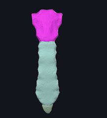
the sternum is the bony structure that makes the thoracic cage from the anterior side, it is also the place where the ribs will be attached to, by the costal cartilage, the costal cartilages are part that is located at the end of the ribs, so we can say that the ribs are indirectly connected to the sternum via the costal cartilages, back to the sternum u can see the photo of the sternum below.
So the sternum is made up of three main parts, which are the
1- the manubrium
the manubrium is the upper part of the sternum, it has the jugular notch, where we can know the place of the clavicle, it is also at the level of the inferior border of the second thoracic vertebra, it is also where u can palpate the trachea, the manubrium on the sides have the places where the first rib and part of the second rib will be attached to.
2- the body of the sternum
Formed from the fusion of four segments (sternebrae). Transverse ridges on the anterior surface
indicate the lines of fusion of the sternebrae. Incomplete ossification of the sternebrae may be shown as a hole in the sternum, called the sternal foramen.
3- the xiphoid process :
is located in the infrasternal subcostal handle, is in the level of the 10th vertebra, and it usually calcifies after the age of 40y, as it will be felt as a hard lump in the epigastrium
with this, we finished the first part of the anatomy of the thoracic cage


.jpg)

Comments
Post a Comment